Xanthélasma is a common condition that causes aesthetic discomfort and does not decrease with age. It is a xanthome plan palpébral (an infiltrate of histiocytic or macrophage cells filled with lipids – esterified cholesterol) that forms a yellowish plaque which gradually extends and darkens over the years on the upper or lower eyelid.
The best treatment is surgical, performed by an ophthalmologist, or if the lesions are small, they can be removed with a laser by a dermatologist.
However, recurrences are common, around 50%, which necessitates further treatments.
The major issue is the risk of ectropion due to skin retraction after multiple treatments.
Ectropion refers to the outward turning of the eyelid's free edge ("eversion" of the eyelid), causing a loss of contact between the eye and the eyelid.
The causes can be diverse:
Ectropion involutif: occurring due to tissue changes (progressive laxity) related to aging.
Ectropion paralytique: secondary to facial paralysis, related to certain myopathies, etc.
Cicatricial entropion: following burns or certain diseases (such as trachoma, etc.).
The treatment for ectropion is surgical and involves the correct repositioning of the eyelid tissues.
Le ptérygion est une tumeur bénigne d’origine conjonctivale envahissant la cornée. Il est le plus souvent situé au niveau du « blanc » de l’œil dans l’angle interne des paupières (le canthus), du côté du nez. Cette tumeur conjonctivo-élastique adopte le plus souvent la forme d’un triangle comparé à une « aile » (étymologie grecque du terme pterys).
Favorisé par l’exposition aux UV et la sécheresse oculaire.
Le traitement chirurgical du ptérygion comporte plusieurs objectifs :
Garder la fonction visuelle menacée par l’envahissement progressif de la surface cornéenne, restaurer l’esthétique de l’œil, éviter les récidives, restaurer la fonction visuelle en cas d’envahissement de l’aire pupillaire.
Spontanément, après une exérèse simple la récidive survient en effet dans une proportion importante des cas, variable selon l’évolution de la lésion et le caractère primaire ou secondaire de l’intervention.
Il faut bien poser le moment de l’indication opératoire car plus on opère, plus le risque de récidive augmente.
Cela pose des problèmes thérapeutiques, du fait de l’épuisement du capital conjonctival disponible pour l’autogreffe. Il est donc capital de mettre en œuvre la meilleure stratégie préventive possible dès la première intervention de ptérygion.
Pour éviter les récidives fréquentes, certaines méthodes sont basées sur la destruction des cellules fibroblastiques conjonctivales application de mitomycine C ou d’autre agent antimétabolite (thiothépa), radiothérapie par irradiation bêta au Strontium 90.
La majorité des méthodes actuellement employées font une place à la reconstruction de la zone d’exérèse :
>par une autogreffe de conjonctive, excellente méthode et simple,
Ptosis refers to a drooping of the upper eyelid. It can be unilateral or bilateral, depending on whether it affects one eye or both, and is caused by a deficiency of the "levator" muscle of the upper eyelid.
Several factors can be the cause of ptosis:
In the case of aging, the eyelid muscle thins and may sag with age.
A neuromuscular disorder, ptosis can then be the first sign of a muscle disease; trauma, such as a blow to the eyebrow arch; or a congenital predisposition.
The main treatment is based on a simple surgical procedure. It involves "re-attaching" the upper eyelid to the muscle. Health insurance will not cover this procedure if the purpose is solely aesthetic.
Very fine and delicate, blepharoplasty (correction of the lower and/or upper eyelids) provides great satisfaction by rejuvenating the eye contour.
Most often performed under local anesthesia with or without sedation, but sometimes upon request, under general anesthesia. The procedure lasts about one hour, and you can leave after one to two hours of rest or stay overnight.
The excess skin and fat pockets are removed through an incision in the natural fold, at the crease of the upper eyelid. The fat pockets of the lower eyelid can be removed through an incision beneath the lash line if there is excess skin. Otherwise, this can also be done without a scar via conjunctival approach. A bandage is optional for a few hours to avoid hematomas and bruising.
You may have traces of the procedure for 8 to 10 days, but activity is still possible with sunglasses with tinted lenses. It is a delicate and meticulous procedure, but it is light and has nothing to do with the ocular globe or vision.
Complications are exceptional.
An ectropion (outward turning of the lower eyelid) may occur if the technique is not properly performed. The eye contour will be rejuvenated, but a few years later, there may be the possibility of another procedure.
Vision is blurred both at distance and up close. The cornea is thinned and distorted, causing irregular astigmatism and myopia. The image is formed on two different planes.
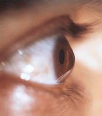
Keratoconus is classified into four stages, and its progression may ultimately lead to a corneal transplant.
The goal of keratoconus treatment is to regularize astigmatism and correct some of the myopia. When keratoconus is advanced, treatment can also delay the need for a corneal transplant or even eliminate the need for this surgery.
The two gold-standard techniques to date are intracorneal rings and Cross-Linking (CXL).
Indications: At stage 1, when the cornea is clear with myopia and astigmatism well corrected by glasses or contact lenses (most often soft or rigid), keratoconus can be treated with the Cross-Linking technique.
At stages 2 and 3, when the cornea is still clear but the astigmatism is too severe to be corrected with glasses (only rigid contact lenses allow for effective vision at these stages), keratoconus can be treated by placing intracorneal rings.
At stage 4, when vision can no longer be corrected with rigid lenses and the cornea shows opacities obstructing vision quality, a corneal transplant is necessary.
>Superficial foreign bodies, which are benign, and intraocular foreign bodies, which pose a risk to visual prognosis.
Injuries can occur simultaneously at different parts of the eye: eyelid, cornea, conjunctiva, iris, capsule, lens, vitreous, and retina. We will not detail all possible injuries, as they are as varied as the nature of the trauma itself.
In all cases, it is essential to consult an Ophthalmology Emergency Service as soon as possible.
The ocular tumors that are more serious include lymphomas, melanomas, and retinoblastomas.
Most often, patients show no symptoms, and the tumor is discovered incidentally during a routine eye examination. When symptoms do occur, the most common symptom of eye cancer is painless vision loss.
Treatment options include:
Treatment options include: chemotherapy, cryotherapy (freezing therapy), enucleation (removal of the eye), laser therapy, observation of slow-growing cancers, radiotherapy, surgery, and thermotherapy (heat therapy).
Possible complications include: recurrence of cancer or its spread, emotional and social difficulties, side effects of treatment, and permanent vision loss.
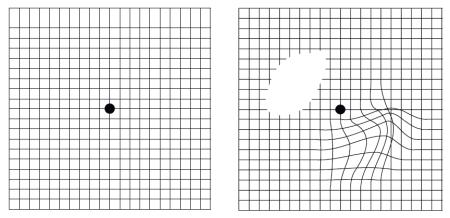
Age-related macular degeneration (AMD) is a disease affecting the eyes which, if left untreated, can lead to rapid sight loss.
The cause is a degeneration of the cells in the central part of the retina called macula. This can lead to a loss of central vision, while leaving peripheral vision usually intact.
There are two forms, the ‘dry’ form and the ‘wet’ form, both of which have the same impact on vision, but develop at different rates. To stop and improve visual acuity, there are treatments using intra vitreous injection.
In industrialised countries, AMD is the leading cause of vision loss in people over 60. The prevalence of AMD increases with age, affecting one in 3 people over 75. Without treatment, half of these people can lose their sight, and for some of them, in just one year!
Withsome adaptations, it is possible to help your eyesight to be protected from AMD, such as: stopping smoking, having your eyesight checked regularly, eating a balanced diet or protecting your eyes from the sun's rays and glare.