The website of your ophthalmologist in Brabant Wallon 

Grand'Place 32

1370 Jodoigne

010 56 00 02

Call us for more information

info@ophtalmo-bw.be

Contact Us by Email, and We'll Reply Soon

Instagram

Follow us on Instagram

Post-Cataract Surgery Care

You are discharged from hospital the same day. You will be given an eye bandage and a protective shell to prevent any eye trauma. These can be removed a few hours later.

Visual acuity returns to normal a few days after the operation. However, this depends on the individual patient and the degree of cataract. The more severe the cataract, the longer it may take to recover.

You will be prescribed treatment for 6 weeks with an antibiotic eye drop and an anti-inflammatory to prevent any inflammation.

Some signs may appear after the operation, but nothing to worry about:

  • The eyes may be red and feel like grains of sand. This is due to scarring and may last a few days,
  • Blurred vision on the day of the operation and the following day is normal,
  • Double vision may occur as a result of the anaesthetic, but this does not last long.

You can resume your daily activities without any problem, but you must remain cautious when it comes to sport, as the eye remains fragile even if vision becomes clear again. For sports involving impact, you should wait 3 weeks. You can wash your hair, but avoid any contact between the eye and water. You can keep or stop wearing your old glasses. If the lenses bother you, you can replace them with neutral lenses.

Decoding a Glasses Prescription

The data is expressed in dioptres.

This is one of the vision tests carried out in the office by your ophthalmologist, who will explain it to you as a measure of visual acuity from 0 to 10 out of 10 (sometimes more in some patients after laser surgery).

  • the first value is the spherical power of the lens. A ‘+’ sign means you are hypermetropic (eye too short or cornea too flat) and a ‘-’ sign means you are myopic (eye too long or cornea too arched), or ‘plane’ there is no correction.
  • The second value is indicated in parentheses. It represents the cylindrical power of the lens, which corresponds to astigmatism (an oval cornea with a flat side on one axis and more curved on the opposite axis of 90°). There is not always a correction. Sometimes expressed in negative (most often by doctors) or in positive (most often by opticians), it makes no difference.
  • The third value is only present to complete the second. It represents the axis of rotation relative to the horizontal, expressed in degrees, and allows for the proper correction of your astigmatism.

Eyelid care - Blepharitis

Blepharitis

Blepharitis is inflammation of the eyelid margins, causing redness, irritation, itching and crusting of the eyelashes. Blepharitis can be caused by microbes or rosacea. It affects people of all ages, but is more common in old age.

The edges of the eyelids are made up of small glands that produce the fat necessary for good quality tears. The fat prevents the tears from evaporating too quickly from the surface of the eye.

In blepharitis, the fat in the glands is rigid and not fluid. It remains blocked, creating inflammation at the edge of the eyelids. The tears do not have enough fat, are of poor quality and evaporate too quickly. The result is dry eyes.

The best way to treat blepharitis is to keep the eyelids clean and free of scabs. It is advisable to stop using make-up.

In the case of a bacterial infection, an antibiotic ointment may also be prescribed, and for dry eyes, your ophthalmologist may prescribe artificial tears.

Treating blepharitis

Start by applying compresses that are as warm as possible for 10 minutes to your closed eyelids. There are also masks that can be heated in the microwave. The aim is to soften the crusts and fluidify the abnormally solid oily secretions contained in the glands

Next, gently massage the upper and lower eyelids, making small circles very close to the eyelashes. From top to bottom for the upper eyelids and from bottom to top for the lower eyelids.

Finally, rinse the eye with saline solution. Finish by caring for the edge of the eyelids with a cleansing wipe or special ointment.

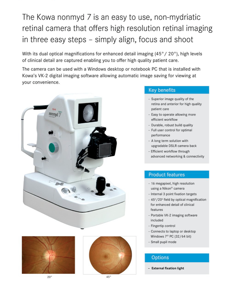
The Jodoigne ophthalmology centre gets a new device to explore the retina

Permettant un meilleur suivi de nos patients, le centre a décidé d’investir et de s’équiper dernièrement d’un rétinographe KOWA 7 (made in USA).

Celui ci permet la réalisation de photographie de la rétine avec une haute précision grâce à une résolution d’image à 16 mégapixels permettant des agrandissements très nets sur des toutes petites surfaces de la rétine invisibles à l’œil nu.

Cet appareil permet en outre de suivre l’évolution du nerf optique, de la macula, de l’état des vaisseaux rétiniens, structures anatomiques altérées dans de nombreuses pathologies comme le glaucome, la DMLA, les thromboses, l’hypertension artérielle et bien sûr le diabète.