The website of your ophthalmologist in Brabant Wallon 

Grand'Place 32

1370 Jodoigne

010 56 00 02

Call us for more information

info@ophtalmo-bw.be

Contact Us by Email, and We'll Reply Soon

How to know if I am a candidate for LASIK?

Step 1: Contact us if you decide that you now want to permanently say goodbye to your glasses and contact lenses.

We will schedule an appointment for a preoperative clinical evaluation. 

You can also do so on our website: under the "Schedule an Appointment" tab / "Ophthalmologist" / "Laser Assessment."

Soit par mail : in**@*********bw.be

For any questions or if you can't find a suitable appointment, you can directly reach the laser secretary at 010/ 56 00 02.

tep 2: Preoperative Clinical Evaluation 

You should plan for 1 hour on-site.

You will be examined, and a complete assessment will be carried out beforehand: vision check, intraocular pressure measurement, retina examination, topography, and pupillometry.

Corneal Topography
Aberrometer, for a unique, personalized treatment for each patient.

We will administer eye drops to dilate your pupils. Please note that this may affect your ability to drive on the way back. We recommend being accompanied.

At the end of the assessment, Dr. Sion will determine if you are a suitable candidate and will guide you toward the technique that best suits you. He will explain everything you need to know to make a clear decision. 

All your questions are important and will be answered. 

The use of soft contact lenses should be stopped at least 48 hours in advance, preferably, and rigid lenses for about 2 weeks.

The basic requirements for laser vision correction include:

1. You would like to stop depending on glasses and contact lenses and enjoy the freedom that this brings.

2. Your prescription is between -1 and -10 diopters (myopia), or up to +5 diopters (hyperopia), with or without a cylinder up to 4 diopters (astigmatism).

3. You are between 20 and 50 years old.

4. You rely on glasses or contact lenses for distance vision.

5. you have stable vision. (If you have your old glasses prescriptions, it is preferable to bring them with you.)

6. you do not suffer from any eye disease and have good vision in both eyes with your glasses.

7. You are in good general health.

Note: Laser vision correction can also help patients who need glasses for both reading and distance vision, but it is not intended for those who have good distance vision and only need glasses for reading.

Step 3: Laser Treatment Day 

The treatments are performed at the Clinique du Parc in Maubeuge and at the Centre Médical Alliance in Braine L’Alleud.

Please present yourself at the reception. You will be re-examined to confirm your prescription and undergo further diagnostic evaluations, followed by the administration of anesthetic eye drops. 

You will be in the laser room with Dr. Sion for about 10 to 15 minutes, while the laser treatment itself takes about 3 to 30 seconds, depending on the correction needed. 

Dr. Sion will provide you with useful information about the application of eye drops.

Overall, you will be on site for about 1 to 2 hours.

100% Laser Treatment: FEMTOSECOND LASER followed by EXCIMER LASER

Stage 4: Follow-up 

All the pre-operative examinations and the operation take place at the Clinique du Parc, with follow-up in Jodoigne.

Patients who have undergone Lasik surgery should be seen again the following day.

PRK patients are generally seen 3 to 5 days after the operation to remove the contact lens. 

Most patients who undergo Lasik surgery are able to resume their activities the next day. 

Most PRK patients can usually return to work after about 5 days with good functional vision. 

In the studies, the final maximum vision was identical at 3 months for both techniques. 

Some post-operative restrictions:

It's important not to rub your eyes or let water get into them for the first week. 

There are no restrictions on reading, using the computer, leaving the house or playing sport. Your eyes may be more sensitive than usual, particularly in the sun or in a smoky atmosphere. 

Sunglasses should be worn outdoors depending on the amount of sunlight, especially after a PRK.

Make-up should be avoided for the first week and care should be taken not to rub the eyes when removing make-up. 

Swimming and opening your eyes underwater should be avoided for 3 weeks. 

Step 5: Make the most of years of glasses-free vision

Post-Laser Vision Correction Care

You will need to be accompanied by car after the laser procedure.

Eye drops should be started as soon as you return home.

After LASIK surgery:

  • The hulls should be kept for 7 nights.
  • A sensation of grains of sand is common, with watery eyes.
  • Above all, don't hesitate to keep your eyes well hydrated with prescribed artificial tears (plenty of them, even 6 to 10 times a day if it makes you feel better).
  • Visual autonomy returns the next day.
  • Healing continues for the first month.

After a PRK operation :

  • A soft lens (conventional, without correction) is fitted after the operation. The aim is to improve healing and prevent excessive eye pain (bandage effect). This lens should be removed between the 3rd and 4th day after the operation.
  • Intense pain and photophobia are common in the first 2 days.
  • Painkillers are prescribed and must be taken immediately to help control the discomfort and pain.
  • Allow for 3 to 5 daysof social downtime (no computer use or driving).
  • Visual autonomy is slower than with LASIK, taking 5 to 7 days.
  • Healing continues for the first 3 months.

Note: At 3 months post-laser, neither technique (LASIK or PRK) has shown superiority in visual outcomes.

Cataract

Cataracte

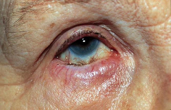
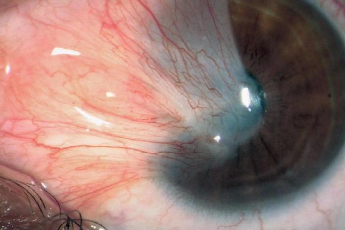
Cataracts correspond to the opacification of the crystalline lens (a normally translucent, high-power lens located inside the eye). Every year, 570,000 people are operated on in France. Surgery is the only way to improve vision altered by cataract.

There are no alternatives to cataract surgery.

Because neither eye drops nor lasers can cure an established cataract. In very advanced cases, cataract can cause blindness.

Worldwide, it is the leading cause of blindness due to the impossibility of large-scale treatment in third world countries. Tens of millions of people are waiting to be operated on, but unfortunately, due to a lack of human and financial resources, many of them will no longer be able to see because of their cataracts. For this reason, many humanitarian trips are organised, particularly to Africa.

Intervention is defined when the opacification of the lens is sufficiently significant, vision deteriorates slowly, often in distance vision with sometimes paradoxically a transient improvement in near vision without glasses.


The degree of discomfort is not the same for everyone. The decision to undergo an operation is therefore the result of a joint agreement between the ophthalmologist and the patient. Without surgery, the cataract will become denser and vision worse, and this may eventually affect fundus examination. Long-term cataract surgery has a current success rate of over 99.5%. 

The surgery:

The procedure is usually performed under local anaesthetic using powerful anaesthetic drops.

Once the eye has been anaesthetised, a micro incision (approximately 2.2 mm) is made at the extreme periphery of the cornea. Then, using a ultrasound probe, the nucleus of the crystalline lens is destructured without damaging the capsule separating it from the posterior part of the eye (this is known as ‘phako-emulsification’). The nucleus of the crystalline lens is then removed to be replaced by a flexible implant. This is inserted folded, then unfolded where the crystalline lens used to be. There are several types of implant.

Currently, the femtosecond laser is used in cataract surgery.

It enables a precise incision to be made, the rhexis, a circular ring around the anterior capsule, to be cut and the nucleus to be cut. The result is greater precision and the use of less ultrasound. Its value is still debated.

In around 1 in 3 cases, a secondary cataract develops, corresponding to an opacification of the posterior capsule (left in deliberately during the operation in order to hold the implant in place). If the patient is bothered by a drop in visual acuity or a sensation of haze, this secondary cataract is treated by laser.

The treatment is carried out in consultation after instillation of drops to dilate the pupil. It is simple, safe, fast (usually less than 10 seconds) and painless.

Dacryocystitis

Une dacryocystite est une inflammation, généralement d’origine infectieuse, d’un sac lacrymal. La dacryocystite se manifeste par un larmoiement continu typique, comme celui que l’on peut connaître lors d’un rhume. Elle survient essentiellement aux âges extrêmes de la vie: chez les nouveau-nés et les personnes âgées de plus de 70 ans. La dacryocystite est liée, chez les personnes âgées, à un rétrécissement des canaux lacrymaux. La baisse d’abondance des larmes entraîne un risque accru d’infection.

Dans les phases débutantes de l’infection, le traitement consistera en une antibiothérapie joint à une application locale d’un collyre antiseptique. 

Lorsque l’infection est plus évoluée, un abcès du sac lacrymal peut survenir, on observe alors l’apparition d’une tuméfaction rouge et douloureuse à la commissure des paupière, la maladie justifie dans ce cas une brève hospitalisation. 

Après de nombreuses récidives et selon la gêne du patient, une dacryo-cysto-rhinostomie peut être effectuée.

La dacryo-cysto-rhinostomie par voie externe est le traitement des obstacles situés sur le trajet du canal lacrymo-nasal. L’intervention est également réalisée par voie endonasale avec des résultats et des complications similaires, hormis l’absence de cicatrice avec cette technique.

Elle consiste à court-circuiter l’obstacle en réalisant une stomie entre le sac lacrymal et la paroi externe de la fosse nasale.

Une anastomose se fera entre le sac lacrymal et la muqueuse des fosses nasales. C’est une intervention préférable à la dacryocystectomie en cas de dacryocystite, parce qu’elle n’entraînera pas un larmoiement permanent pour le patient. L’intervention a un taux de réussite d’environ 90% et fait disparaître tous les symptômes. Elle est cependant plus difficile et plus longue qu’une dacryocystectomie et requiert un matériel et un savoir faire spécifique.

L’intervention consiste à extraire un morceau de la paroi osseuse entre le sac lacrymal et le méat moyen du nez, puis de suturer la muqueuse du sac à la muqueuse nasale du méat moyen.

Xanthelasma

Xanthélasma is a common condition that causes aesthetic discomfort and does not decrease with age. It is a xanthome plan palpébral (an infiltrate of histiocytic or macrophage cells filled with lipids – esterified cholesterol) that forms a yellowish plaque which gradually extends and darkens over the years on the upper or lower eyelid.

The best treatment is surgical, performed by an ophthalmologist, or if the lesions are small, they can be removed with a laser by a dermatologist.

However, recurrences are common, around 50%, which necessitates further treatments.

The major issue is the risk of ectropion due to skin retraction after multiple treatments.

Ectropion

Ectropion refers to the outward turning of the eyelid's free edge ("eversion" of the eyelid), causing a loss of contact between the eye and the eyelid.

The causes can be diverse:

Ectropion involutif: occurring due to tissue changes (progressive laxity) related to aging.

Ectropion paralytique: secondary to facial paralysis, related to certain myopathies, etc.

Cicatricial entropion: following burns or certain diseases (such as trachoma, etc.).

The treatment for ectropion is surgical and involves the correct repositioning of the eyelid tissues.

Entropion

Entropion refers to the inward turning of the eyelid's free edge, causing contact between the eye and the eyelashes.

The causes can be diverse:

Spasmodic entropion (for example, following a surgical intervention)

Involutive entropion: occurring due to tissue changes (progressive laxity) related to aging.

Cicatricial entropion: following burns or certain diseases (such as trachoma, etc.).

Congenital entropion: present at birth.

The treatment for entropion is surgical and involves the correct repositioning of the eyelid tissues.

Pterygium

http://www.zioneye.com/eye-surgery/reconstructive-and-cosmetic-eyelid-surgery/pterygium/

Le ptérygion est une tumeur bénigne d’origine conjonctivale envahissant la cornée.
Il est le plus souvent situé au niveau du « blanc » de l’œil dans l’angle interne des paupières (le canthus), du côté du nez.
Cette tumeur conjonctivo-élastique adopte le plus souvent la forme d’un triangle comparé à une « aile » (étymologie grecque du terme pterys).

Favorisé par l’exposition aux UV et la sécheresse oculaire.

Le traitement chirurgical du ptérygion comporte plusieurs objectifs :

Garder la fonction visuelle menacée par l’envahissement progressif de la surface cornéenne, restaurer l’esthétique de l’œil,  éviter les récidives, restaurer la fonction visuelle en cas d’envahissement de l’aire pupillaire.

Spontanément, après une exérèse simple la récidive survient en effet dans une proportion importante des cas, variable selon l’évolution de la lésion et le caractère primaire ou secondaire de l’intervention. 

Il faut bien poser le moment de l’indication opératoire car plus on opère, plus le risque de récidive augmente.

Cela pose des problèmes thérapeutiques, du fait de l’épuisement du capital conjonctival disponible pour l’autogreffe.
Il est donc capital de mettre en œuvre la meilleure stratégie préventive possible dès la première intervention de ptérygion.

Pour éviter les récidives fréquentes, certaines méthodes sont basées sur la destruction des cellules fibroblastiques conjonctivales application de mitomycine C ou d’autre agent antimétabolite (thiothépa), radiothérapie par irradiation bêta au Strontium 90.

La majorité des méthodes actuellement employées font une place à la reconstruction de la zone d’exérèse :

>par une autogreffe de conjonctive, excellente méthode et simple,

>par une autogreffe cornéenne,

>par un allogreffe cornéenne lamellaire.

Ptosis

Ptosis refers to a drooping of the upper eyelid. It can be unilateral or bilateral, depending on whether it affects one eye or both, and is caused by a deficiency of the "levator" muscle of the upper eyelid.

Several factors can be the cause of ptosis: 

In the case of aging, the eyelid muscle thins and may sag with age. 

A neuromuscular disorder, ptosis can then be the first sign of a muscle disease; trauma, such as a blow to the eyebrow arch; or a congenital predisposition.

The main treatment is based on a simple surgical procedure. It involves "re-attaching" the upper eyelid to the muscle. Health insurance will not cover this procedure if the purpose is solely aesthetic.