The website of your ophthalmologist in Brabant Wallon 

Grand'Place 32

1370 Jodoigne

010 56 00 02

Call us for more information

info@ophtalmo-bw.be

Contact Us by Email, and We'll Reply Soon

Instagram

Follow us on Instagram

Chalazion

The consequence of a chalazion is inflammation caused by the blockage of the drainage duct of a Meibomian gland, located inside the upper and/or lower eyelid.

This then manifests as a small bump on the eyelid, which can grow in size and may become red, painful, with a sensation of heat over the course of several days.
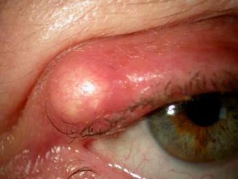
Typically, a chalazion disappears after a few weeks, and in this case, there are no medical consequences. A chalazion is not contagious.

Stye and chalazion

Even though a stye is also a bump in the eyelid caused by the obstruction of a sebaceous gland, a chalazion is not a stye. A stye represents an acute infection of the gland. A chalazion is not an infection but rather an inflammation of the area. Inflammation is a process in which the body reacts to a condition and produces swelling, redness, pain, or heat. A stye is usually more painful than a chalazion and may appear infected.

A chalazion is caused by the oil from the Meibomian gland becoming too thick to flow out of the gland. The oil then accumulates within the gland, forming a bump in the eyelid. The gland's wall may rupture, releasing the oil into the tissue of the eyelid, which then causes inflammation of the tissues and sometimes even scarring.

Medical Treatment

The treatment for a chalazion may include the following:

Applying warm compresses for five to ten minutes, four times a day, to reduce swelling and encourage drainage of the gland.

A medical prescription for antibiotic drops or ointments if a bacterial infection is suspected.

Rarely, an injection of a steroid medication into the area of the bump (the chalazion) to help reduce inflammation.

If a chalazion causes significant symptoms or lasts for several weeks, it may be necessary to remove it surgically. If the appearance of the chalazion bothers the patient, surgical removal may also be performed.

In the case of multiple chalazia or recurrent chalazia, the possibility that the patient may have a generalized condition affecting the sebaceous glands should be considered. This is the case with rosacea: this condition can be treated long-term with oral medication to alter the consistency of the oils produced by the glands.

Dacryocystitis

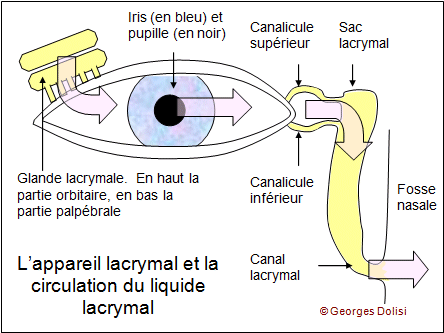
Une dacryocystite est une inflammation, généralement d’origine infectieuse, d’un sac lacrymal. La dacryocystite se manifeste par un larmoiement continu typique, comme celui que l’on peut connaître lors d’un rhume. Elle survient essentiellement aux âges extrêmes de la vie: chez les nouveau-nés et les personnes âgées de plus de 70 ans. La dacryocystite est liée, chez les personnes âgées, à un rétrécissement des canaux lacrymaux. La baisse d’abondance des larmes entraîne un risque accru d’infection.

Dans les phases débutantes de l’infection, le traitement consistera en une antibiothérapie joint à une application locale d’un collyre antiseptique. 

Lorsque l’infection est plus évoluée, un abcès du sac lacrymal peut survenir, on observe alors l’apparition d’une tuméfaction rouge et douloureuse à la commissure des paupière, la maladie justifie dans ce cas une brève hospitalisation. 

Après de nombreuses récidives et selon la gêne du patient, une dacryo-cysto-rhinostomie peut être effectuée.

La dacryo-cysto-rhinostomie par voie externe est le traitement des obstacles situés sur le trajet du canal lacrymo-nasal. L’intervention est également réalisée par voie endonasale avec des résultats et des complications similaires, hormis l’absence de cicatrice avec cette technique.

Elle consiste à court-circuiter l’obstacle en réalisant une stomie entre le sac lacrymal et la paroi externe de la fosse nasale.

Une anastomose se fera entre le sac lacrymal et la muqueuse des fosses nasales. C’est une intervention préférable à la dacryocystectomie en cas de dacryocystite, parce qu’elle n’entraînera pas un larmoiement permanent pour le patient. L’intervention a un taux de réussite d’environ 90% et fait disparaître tous les symptômes. Elle est cependant plus difficile et plus longue qu’une dacryocystectomie et requiert un matériel et un savoir faire spécifique.

L’intervention consiste à extraire un morceau de la paroi osseuse entre le sac lacrymal et le méat moyen du nez, puis de suturer la muqueuse du sac à la muqueuse nasale du méat moyen.

Xanthelasma

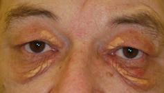
Xanthélasma is a common condition that causes aesthetic discomfort and does not decrease with age. It is a xanthome plan palpébral (an infiltrate of histiocytic or macrophage cells filled with lipids – esterified cholesterol) that forms a yellowish plaque which gradually extends and darkens over the years on the upper or lower eyelid.

The best treatment is surgical, performed by an ophthalmologist, or if the lesions are small, they can be removed with a laser by a dermatologist.

However, recurrences are common, around 50%, which necessitates further treatments.

The major issue is the risk of ectropion due to skin retraction after multiple treatments.

Ectropion

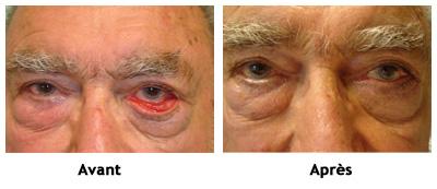
Ectropion refers to the outward turning of the eyelid's free edge ("eversion" of the eyelid), causing a loss of contact between the eye and the eyelid.

The causes can be diverse:

Ectropion involutif: occurring due to tissue changes (progressive laxity) related to aging.

Ectropion paralytique: secondary to facial paralysis, related to certain myopathies, etc.

Cicatricial entropion: following burns or certain diseases (such as trachoma, etc.).

The treatment for ectropion is surgical and involves the correct repositioning of the eyelid tissues.

Entropion

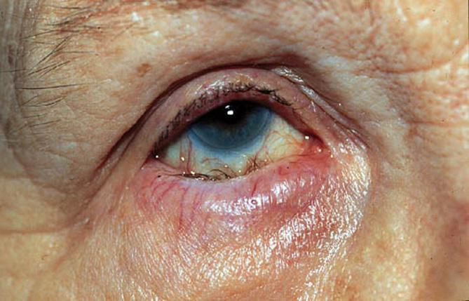
Entropion refers to the inward turning of the eyelid's free edge, causing contact between the eye and the eyelashes.

The causes can be diverse:

Spasmodic entropion (for example, following a surgical intervention)

Involutive entropion: occurring due to tissue changes (progressive laxity) related to aging.

Cicatricial entropion: following burns or certain diseases (such as trachoma, etc.).

Congenital entropion: present at birth.

The treatment for entropion is surgical and involves the correct repositioning of the eyelid tissues.

Pterygium

http://www.zioneye.com/eye-surgery/reconstructive-and-cosmetic-eyelid-surgery/pterygium/

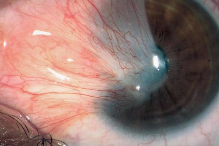
Le ptérygion est une tumeur bénigne d’origine conjonctivale envahissant la cornée.
Il est le plus souvent situé au niveau du « blanc » de l’œil dans l’angle interne des paupières (le canthus), du côté du nez.
Cette tumeur conjonctivo-élastique adopte le plus souvent la forme d’un triangle comparé à une « aile » (étymologie grecque du terme pterys).

Favorisé par l’exposition aux UV et la sécheresse oculaire.

Le traitement chirurgical du ptérygion comporte plusieurs objectifs :

Garder la fonction visuelle menacée par l’envahissement progressif de la surface cornéenne, restaurer l’esthétique de l’œil,  éviter les récidives, restaurer la fonction visuelle en cas d’envahissement de l’aire pupillaire.

Spontanément, après une exérèse simple la récidive survient en effet dans une proportion importante des cas, variable selon l’évolution de la lésion et le caractère primaire ou secondaire de l’intervention. 

Il faut bien poser le moment de l’indication opératoire car plus on opère, plus le risque de récidive augmente.

Cela pose des problèmes thérapeutiques, du fait de l’épuisement du capital conjonctival disponible pour l’autogreffe.
Il est donc capital de mettre en œuvre la meilleure stratégie préventive possible dès la première intervention de ptérygion.

Pour éviter les récidives fréquentes, certaines méthodes sont basées sur la destruction des cellules fibroblastiques conjonctivales application de mitomycine C ou d’autre agent antimétabolite (thiothépa), radiothérapie par irradiation bêta au Strontium 90.

La majorité des méthodes actuellement employées font une place à la reconstruction de la zone d’exérèse :

>par une autogreffe de conjonctive, excellente méthode et simple,

>par une autogreffe cornéenne,

>par un allogreffe cornéenne lamellaire.

Ptosis

Ptosis refers to a drooping of the upper eyelid. It can be unilateral or bilateral, depending on whether it affects one eye or both, and is caused by a deficiency of the "levator" muscle of the upper eyelid.

Several factors can be the cause of ptosis: 

In the case of aging, the eyelid muscle thins and may sag with age. 

A neuromuscular disorder, ptosis can then be the first sign of a muscle disease; trauma, such as a blow to the eyebrow arch; or a congenital predisposition.

The main treatment is based on a simple surgical procedure. It involves "re-attaching" the upper eyelid to the muscle. Health insurance will not cover this procedure if the purpose is solely aesthetic.

Blepharochalasis

Treatment: Blepharoplasty

Very fine and delicate, blepharoplasty (correction of the lower and/or upper eyelids) provides great satisfaction by rejuvenating the eye contour. 

Most often performed under local anesthesia with or without sedation, but sometimes upon request, under general anesthesia. The procedure lasts about one hour, and you can leave after one to two hours of rest or stay overnight.

The excess skin and fat pockets are removed through an incision in the natural fold, at the crease of the upper eyelid. The fat pockets of the lower eyelid can be removed through an incision beneath the lash line if there is excess skin. Otherwise, this can also be done without a scar via conjunctival approach. A bandage is optional for a few hours to avoid hematomas and bruising.

You may have traces of the procedure for 8 to 10 days, but activity is still possible with sunglasses with tinted lenses.
It is a delicate and meticulous procedure, but it is light and has nothing to do with the ocular globe or vision.

Complications are exceptional.

An ectropion (outward turning of the lower eyelid) may occur if the technique is not properly performed.
The eye contour will be rejuvenated, but a few years later, there may be the possibility of another procedure.