The website of your ophthalmologist in Brabant Wallon 

Grand'Place 32

1370 Jodoigne

010 56 00 02

Call us for more information

info@ophtalmo-bw.be

Contact Us by Email, and We'll Reply Soon

Instagram

Follow us on Instagram

Cataract

Cataracte

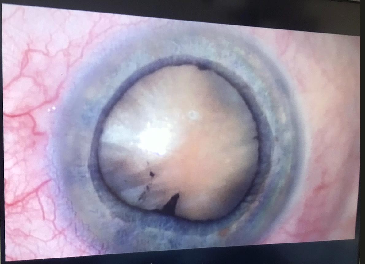
Cataracts correspond to the opacification of the crystalline lens (a normally translucent, high-power lens located inside the eye). Every year, 570,000 people are operated on in France. Surgery is the only way to improve vision altered by cataract.

There are no alternatives to cataract surgery.

Because neither eye drops nor lasers can cure an established cataract. In very advanced cases, cataract can cause blindness.

Worldwide, it is the leading cause of blindness due to the impossibility of large-scale treatment in third world countries. Tens of millions of people are waiting to be operated on, but unfortunately, due to a lack of human and financial resources, many of them will no longer be able to see because of their cataracts. For this reason, many humanitarian trips are organised, particularly to Africa.

Intervention is defined when the opacification of the lens is sufficiently significant, vision deteriorates slowly, often in distance vision with sometimes paradoxically a transient improvement in near vision without glasses.


The degree of discomfort is not the same for everyone. The decision to undergo an operation is therefore the result of a joint agreement between the ophthalmologist and the patient. Without surgery, the cataract will become denser and vision worse, and this may eventually affect fundus examination. Long-term cataract surgery has a current success rate of over 99.5%. 

The surgery:

The procedure is usually performed under local anaesthetic using powerful anaesthetic drops.

Once the eye has been anaesthetised, a micro incision (approximately 2.2 mm) is made at the extreme periphery of the cornea. Then, using a ultrasound probe, the nucleus of the crystalline lens is destructured without damaging the capsule separating it from the posterior part of the eye (this is known as ‘phako-emulsification’). The nucleus of the crystalline lens is then removed to be replaced by a flexible implant. This is inserted folded, then unfolded where the crystalline lens used to be. There are several types of implant.

Currently, the femtosecond laser is used in cataract surgery.

It enables a precise incision to be made, the rhexis, a circular ring around the anterior capsule, to be cut and the nucleus to be cut. The result is greater precision and the use of less ultrasound. Its value is still debated.

In around 1 in 3 cases, a secondary cataract develops, corresponding to an opacification of the posterior capsule (left in deliberately during the operation in order to hold the implant in place). If the patient is bothered by a drop in visual acuity or a sensation of haze, this secondary cataract is treated by laser.

The treatment is carried out in consultation after instillation of drops to dilate the pupil. It is simple, safe, fast (usually less than 10 seconds) and painless.

Keratoconus

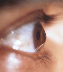
Vision is blurred both at distance and up close. The cornea is thinned and distorted, causing irregular astigmatism and myopia. The image is formed on two different planes.

Keratoconus is classified into four stages, and its progression may ultimately lead to a corneal transplant.

The goal of keratoconus treatment is to regularize astigmatism and correct some of the myopia. When keratoconus is advanced, treatment can also delay the need for a corneal transplant or even eliminate the need for this surgery.

The two gold-standard techniques to date are intracorneal rings and Cross-Linking (CXL).

Indications:
At stage 1, when the cornea is clear with myopia and astigmatism well corrected by glasses or contact lenses (most often soft or rigid), keratoconus can be treated with the Cross-Linking technique.

At stages 2 and 3, when the cornea is still clear but the astigmatism is too severe to be corrected with glasses (only rigid contact lenses allow for effective vision at these stages), keratoconus can be treated by placing intracorneal rings.

At stage 4, when vision can no longer be corrected with rigid lenses and the cornea shows opacities obstructing vision quality, a corneal transplant is necessary.

Ocular trauma

Ocular globe trauma can take many forms.

There are three main types of trauma:

>Contusions du globe (e.g., champagne cork)

>Perforating traumas (wooden branch, nail, arrow)

>Superficial foreign bodies, which are benign, and intraocular foreign bodies, which pose a risk to visual prognosis.

Injuries can occur simultaneously at different parts of the eye: eyelid, cornea, conjunctiva, iris, capsule, lens, vitreous, and retina. We will not detail all possible injuries, as they are as varied as the nature of the trauma itself.

In all cases, it is essential to consult an Ophthalmology Emergency Service as soon as possible.

Ocular tumours

The ocular tumors that are more serious include lymphomas, melanomas, and retinoblastomas.

Most often, patients show no symptoms, and the tumor is discovered incidentally during a routine eye examination. When symptoms do occur, the most common symptom of eye cancer is painless vision loss.

Treatment options include:

Treatment options include: chemotherapy, cryotherapy (freezing therapy), enucleation (removal of the eye), laser therapy, observation of slow-growing cancers, radiotherapy, surgery, and thermotherapy (heat therapy).

Possible complications include: recurrence of cancer or its spread, emotional and social difficulties, side effects of treatment, and permanent vision loss.

AMD (Age-related macular degeneration)

Age-related macular degeneration (AMD) is a disease affecting the eyes which, if left untreated, can lead to rapid sight loss.

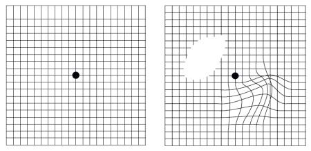
The cause is a degeneration of the cells in the central part of the retina called macula. This can lead to a loss of central vision, while leaving peripheral vision usually intact.

There are two forms, the ‘dry’ form and the ‘wet’ form, both of which have the same impact on vision, but develop at different rates. To stop and improve visual acuity, there are treatments using intra vitreous injection.

In industrialised countries, AMD is the leading cause of vision loss in people over 60. The prevalence of AMD increases with age, affecting one in 3 people over 75. Without treatment, half of these people can lose their sight, and for some of them, in just one year! 

Withsome adaptations, it is possible to help your eyesight to be protected from AMD, such as: stopping smoking, having your eyesight checked regularly, eating a balanced diet or protecting your eyes from the sun's rays and glare.

Glaucoma

Glaucoma is a common eye disease, affecting more than 2% of the population over the age of 45.

In France, it is estimated that approximately 1 million people are affected. This high prevalence justifies regular intraocular pressure measurement during an eye examination.

It is advisable to see your specialist every two years or so (from the age of 45) to have these measurements carried out.

In developed countries, glaucoma is the leading cause of absolute blindness. Other common causes of vision loss include age-related macular degeneration and cataracts. These two conditions are not related to glaucoma but may coexist in patients with glaucoma.

Two major forms of glaucoma exist.

These are open angle glaucoma, the most common, and angle closure glaucoma.

Glaucoma is due to an increase in eye pressure leading to damage to the optic nerve (which sends visual information to the brain) and the visual field (vision space).

Open-angle glaucoma is linked to the progressive closure of the evacuation filter, the trabecular meshwork. This phenomenon is progressive because the patient does not feel the slow increase in eye pressure.

Open-angle glaucoma (OCT examination)

Source: French Glaucoma Society – 2011

Angle-closure glaucoma, also known as acute glaucoma, is a sudden and severe condition. It occurs when the iris quickly adheres to the drainage filter, leading to significant pain and vision loss. This is a major ophthalmologic emergency.

Angle-closure glaucoma (OCT examination)

Source: French Glaucoma Society – 2011

There are also very rare types of glaucoma that occur from birth, called congenital glaucomas. From the outset, their treatment is surgical.

Finally, glaucoma can develop as a result of another eye condition, such as inflammation or trauma..